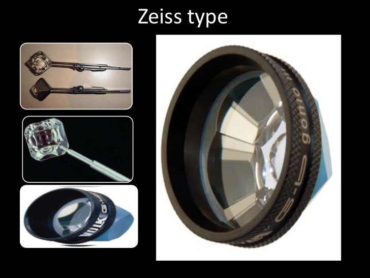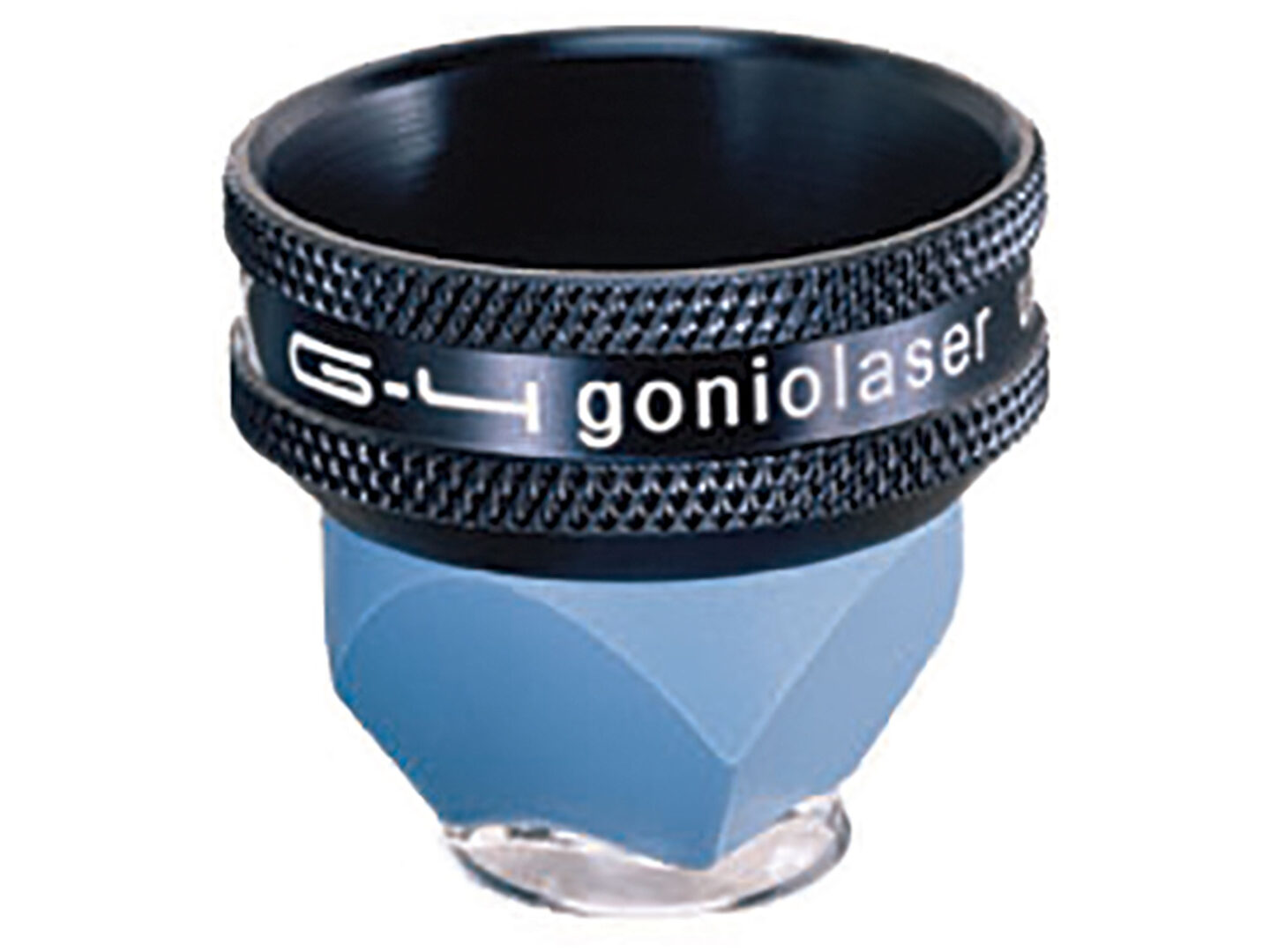
The Zeiss 4 Mirror Gonio Lens is an essential tool in ophthalmology. It allows clinicians to visualize the angle of the anterior chamber. This angle is crucial for diagnosing and managing glaucoma.
Understanding Gonioscopy
Gonioscopy uses a specialized lens. This lens overcomes total internal reflection. It enables direct visualization of the angle structures.
Without a gonio lens, light reflects internally within the eye. The angle structures cannot be seen directly. The lens acts as a refractive intermediary. It allows light to exit the eye at a different angle, providing a clear view.
The Zeiss 4 Mirror Gonio Lens offers a wide field of view. The image is viewed directly. It is often used for quick screening and detailed examination of the iridocorneal angle.
Key Components of the Angle
Understanding angle anatomy is vital. Students should be familiar with key structures. These include the Schwalbe's line, trabecular meshwork, scleral spur, and ciliary body band.
Schwalbe's line is the most anterior structure. It represents the termination of Descemet's membrane. Identifying it is crucial for orienting yourself within the angle.
The trabecular meshwork is posterior to Schwalbe's line. It is responsible for aqueous outflow. Its appearance can indicate various types of glaucoma. The degree of pigmentation within this structure also has diagnostic implications.
The scleral spur is posterior to the trabecular meshwork. It appears as a whitish band. It is an important landmark for identifying the ciliary body band.
The ciliary body band is the most posterior structure. It can vary in width and pigmentation. Its visibility can be affected by the iris insertion.
Teaching Tips for Educators
Start with the basics of angle anatomy. Use diagrams and illustrations to clearly depict the structures. Make use of digital tools to show how the gonio lens modifies the path of light.
Employ interactive simulations. These help students understand the principles of gonioscopy. Consider using virtual reality or augmented reality if accessible. These tools can create an immersive learning experience.
Provide real-world examples of gonioscopy findings. Show images of normal and abnormal angles. Discuss the clinical significance of different findings. Students can then apply their knowledge to solve case studies.
Consider organizing a practical demonstration. Allow students to practice using a gonio lens on a model eye. This hands-on experience will solidify their understanding. Practice on a real patient is best but always requires supervision.
Incorporate case studies that present different types of glaucoma. Ask students to interpret the gonioscopy findings. Encourage critical thinking and problem-solving skills.
Addressing Common Misconceptions
One common misconception is that the gonio lens magnifies the angle structures. Actually, the lens corrects the angle of refraction, making the angle visible. The lens does not increase magnification.
Another misconception is that gonioscopy is only for diagnosing glaucoma. It is also useful for managing glaucoma. Furthermore it can be used for detecting other anterior segment abnormalities.
Some students may struggle to differentiate between angle structures. Emphasize the unique features of each structure. Use mnemonics or visual aids to help students remember the order and appearance. Regularly test their understanding.
Making the Concept Engaging
Use relatable analogies. Explain how the gonio lens works like a periscope. Explain how it allows you to see around a corner, the corner in this instance being the iris.
Incorporate technology. Utilize online resources, videos, and interactive simulations. These will keep students engaged and motivated. Show clinical videos of gonioscopy examinations.
Invite guest speakers. Have experienced ophthalmologists share their experiences with gonioscopy. This can provide valuable insights into the clinical applications of the technique. Encourage students to ask questions and participate in discussions.
Create a game or quiz. This can be a fun way to review the material. Offer prizes for the top performers to increase engagement. Turn lessons into interactive games or challenges. For example, simulate angle closure scenarios and have students predict the gonioscopic appearance.
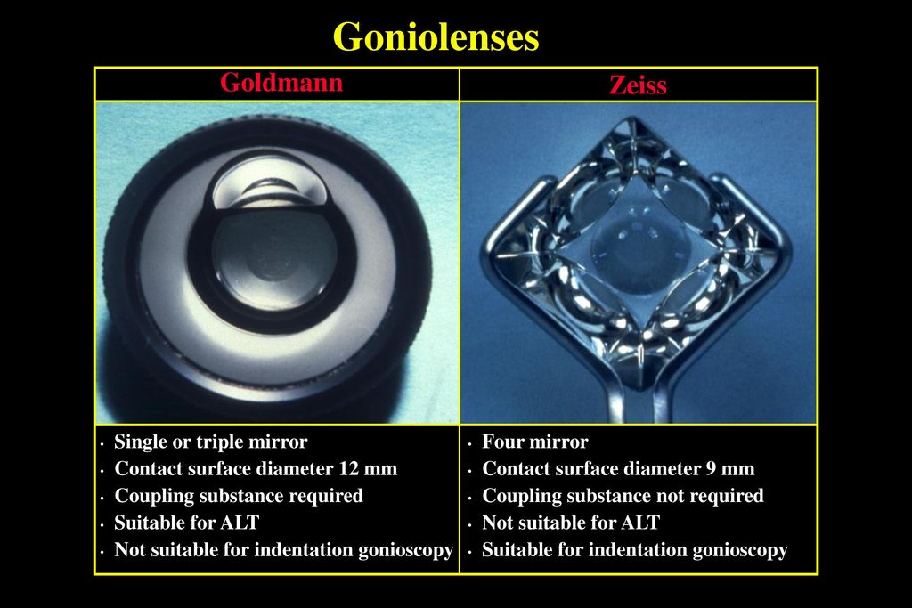
Encourage students to research different types of gonio lenses. They should research the advantages and disadvantages of each. This will promote independent learning and critical thinking.
Focus on the clinical relevance of gonioscopy. Explain how it helps to prevent vision loss. Emphasize the importance of accurate diagnosis and management of glaucoma. Connect theoretical knowledge to practical applications.
Practical Considerations
Before performing gonioscopy, ensure that the patient is comfortable. Explain the procedure to them thoroughly. Address any concerns or questions they may have.
Use topical anesthesia. This will minimize discomfort during the examination. Select the appropriate size gonio lens for the patient. Different lenses are available for different eye sizes and anatomical features.
Apply coupling solution to the lens. This will create a smooth optical interface between the lens and the cornea. Ensure that there are no air bubbles. These can obstruct the view.
Use gentle pressure when placing the lens on the eye. Avoid applying excessive pressure. This can distort the angle structures. Rotate the lens to view all quadrants of the angle. Document your findings in a clear and concise manner.
Proper disinfection is crucial after each use. Follow the manufacturer's instructions for cleaning and sterilizing the lens. This will prevent the spread of infection. It will also ensure the longevity of the lens.
By understanding the Zeiss 4 Mirror Gonio Lens and its applications, students can develop the skills necessary for effective glaucoma management. They can contribute to the prevention of blindness. This knowledge can be invaluable in their future careers.