Understanding the events at the neuromuscular junction (NMJ) is crucial. It bridges the nervous system and skeletal muscles.
The Neuromuscular Junction: A Key Connection
The NMJ facilitates muscle contraction. Nerve impulses translate into muscle action here.
Components of the NMJ
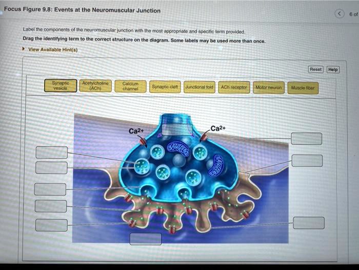
Several structures make up the NMJ. These include the motor neuron, synaptic cleft, and motor end plate.
The motor neuron is a nerve cell. It transmits signals from the brain or spinal cord.
The synaptic cleft is a small gap. It separates the motor neuron and muscle fiber.
The motor end plate is a specialized region. It is on the muscle fiber's plasma membrane.
The Sequence of Events
A series of steps lead to muscle contraction. Understanding these steps is essential.
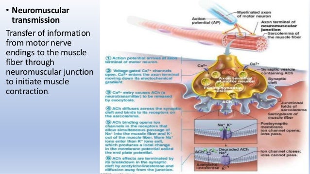
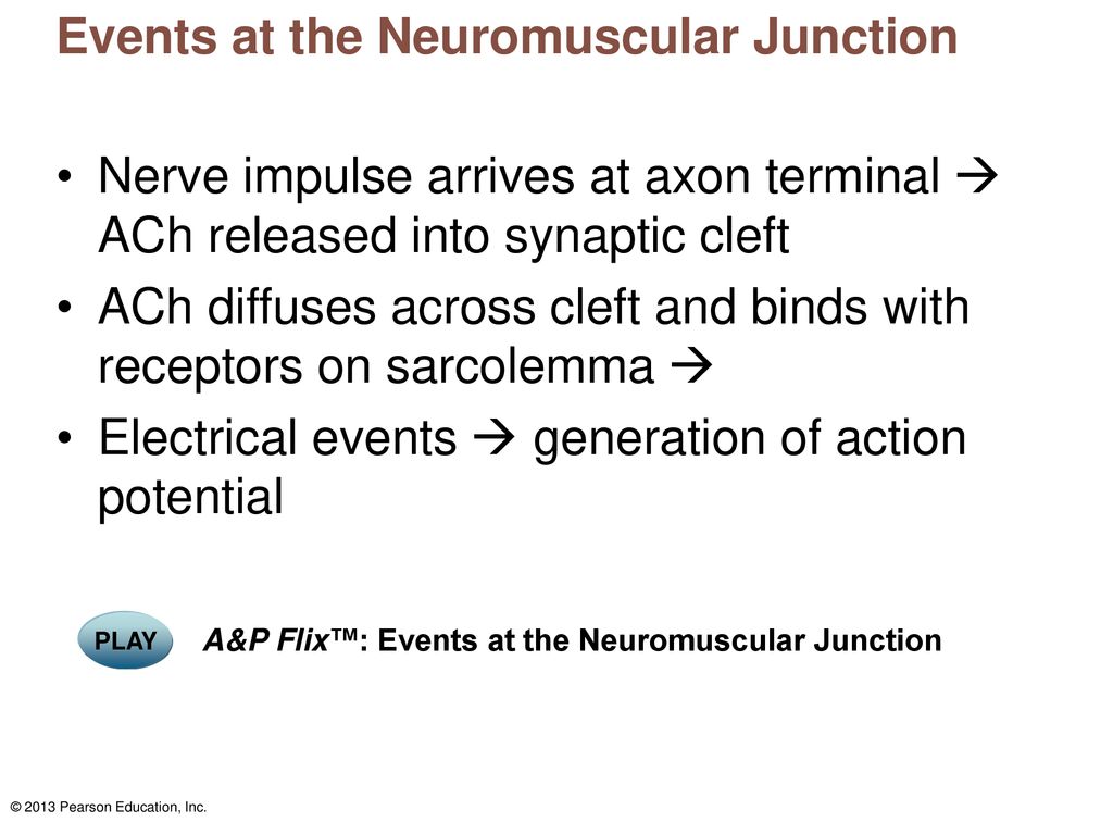
Step 1: Action Potential Arrival. An action potential reaches the motor neuron's axon terminal.
Step 2: Calcium Influx. This depolarization opens voltage-gated calcium channels. Calcium ions (Ca2+) rush into the axon terminal.
Step 3: Acetylcholine Release. The influx of Ca2+ triggers the fusion. Synaptic vesicles fuse with the presynaptic membrane.
Step 4: Acetylcholine Diffusion. Acetylcholine (ACh) diffuses. It travels across the synaptic cleft.
Step 5: Acetylcholine Binding. ACh binds to ACh receptors. These receptors are on the motor end plate.
Step 6: Motor End Plate Depolarization. ACh binding opens ligand-gated ion channels. Sodium ions (Na+) flow into the muscle fiber. Potassium ions (K+) flow out.
Step 7: Action Potential Generation. The influx of Na+ depolarizes the motor end plate. This creates an end-plate potential (EPP).
Step 8: Muscle Fiber Action Potential. If the EPP reaches threshold, an action potential is generated. It propagates along the muscle fiber.
Step 9: Acetylcholine Degradation. Acetylcholinesterase (AChE) breaks down ACh. This prevents continuous muscle stimulation.
Teaching Tips for Educators
Use visual aids. Diagrams and animations can help. They illustrate the events at the NMJ.
Break down the process. Divide it into smaller, manageable steps.
Relate the NMJ to real-world examples. Discuss conditions like myasthenia gravis. This autoimmune disease affects ACh receptors.
Use analogies. Compare the NMJ to a relay race. One runner passes the baton to another.
Incorporate active learning. Have students act out the steps at the NMJ.
Common Misconceptions
Students often confuse the action potential in the neuron. They also confuse it with the action potential in the muscle fiber. Clarify that these are separate events.
Some students think ACh is constantly released. Emphasize that ACh release is triggered. It requires an action potential and calcium influx.
Students might believe that AChE only removes ACh. Explain that it breaks down ACh. This is crucial for muscle relaxation.
Many think that a single action potential. They think it causes a prolonged muscle contraction. Clarify that a single action potential results in a brief twitch.
Making the NMJ Engaging
Use interactive simulations. Virtual labs can help students explore the NMJ.
Create a model NMJ. Use everyday materials like pipe cleaners and beads. Represent different components.
Design a game. A board game or card game. It can reinforce the sequence of events.
Discuss the effects of toxins. Some toxins, like botulinum toxin, affect the NMJ.
Connect the NMJ to exercise physiology. Explain how the NMJ adapts to training. It impacts muscle strength and endurance.
Deep Dive: Acetylcholine's Role
Acetylcholine (ACh) is a neurotransmitter. It plays a crucial role at the NMJ.
ACh is synthesized in the motor neuron's cytoplasm. Choline acetyltransferase catalyzes the reaction.
ACh is then transported into synaptic vesicles. Vesicular acetylcholine transporter (VAChT) does this.
When an action potential arrives. Voltage-gated calcium channels open. Calcium influx triggers ACh release.
ACh diffuses across the synaptic cleft. It binds to nicotinic acetylcholine receptors (nAChRs). These are ligand-gated ion channels.
nAChRs are located on the motor end plate. When ACh binds, the channels open.
Na+ ions flow into the muscle fiber. K+ ions flow out. This creates a depolarization (EPP).
Acetylcholinesterase (AChE) is an enzyme. It breaks down ACh into acetate and choline.
Choline is transported back into the presynaptic terminal. It is used to synthesize more ACh.
Diseases and the NMJ
Several diseases affect the NMJ. Understanding these conditions helps illustrate the NMJ's importance.
Myasthenia Gravis. An autoimmune disorder. Antibodies block or destroy ACh receptors.
Lambert-Eaton Myasthenic Syndrome (LEMS). Antibodies attack voltage-gated calcium channels. This reduces ACh release.
Botulism. Caused by botulinum toxin. It prevents ACh release at the NMJ.
Organophosphate Poisoning. Organophosphates inhibit AChE. This leads to ACh accumulation and overstimulation.
Discussing these diseases. It can provide a clinical context. It highlights the importance of the NMJ.
Conclusion
The neuromuscular junction is a critical site. It is for communication between nerves and muscles.
Understanding the sequence of events. This is crucial for comprehending muscle function.
By using effective teaching strategies. Educators can help students master this important concept.